Trypsin Digestion Protocol for LC–MS/MS (In-Solution)
Learn the complete step-by-step Trypsin Digestion Protocol for LC–MS/MS (In-Solution) including reagent preparation, incubation conditions, enzyme ratios, and troubleshooting tips for high-quality peptide generation in proteomic analysis.
PROTOCOLS
Dr. Mainak Mukhopadhyay
10/29/20254 min read

Introduction
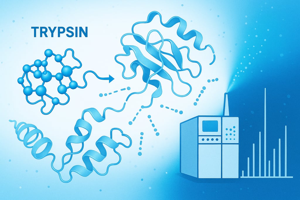
Protein identification and quantification through liquid chromatography–tandem mass spectrometry (LC–MS/MS) has revolutionized proteomics research. One of the most critical steps in this workflow is the enzymatic digestion of proteins into peptides, typically using the protease trypsin.
Trypsin digestion ensures that large, complex proteins are broken down into smaller, charged peptides that are compatible with LC–MS/MS ionization and fragmentation. The quality of peptide generation directly determines the accuracy, reproducibility, and depth of proteomic analysis.
In this detailed guide, we’ll walk through the complete in-solution trypsin digestion protocol for LC–MS/MS sample preparation, explain each step, and highlight important tips for achieving consistent digestion efficiency.
Objective
To enzymatically digest purified protein samples into tryptic peptides suitable for LC–MS/MS analysis, ensuring optimal sequence coverage, reproducibility, and quantification accuracy.
Principle of Trypsin Digestion
Trypsin is a serine protease that specifically cleaves peptide bonds at the C-terminal side of lysine (K) and arginine (R) residues, except when followed by proline. This specificity yields peptides of moderate size (typically 7–25 amino acids), which are ideal for MS detection.
Before digestion, proteins must be:
Denatured to unfold tertiary structures,
Reduced to break disulfide bonds,
Alkylated to prevent reformation of disulfide bonds, and then
Diluted and digested with trypsin under mild, buffered conditions.
This sequential treatment ensures complete and reproducible digestion of complex protein mixtures.
Reagents and Chemicals Required
Step-by-Step Protocol (In-Solution Trypsin Digestion)
1. Denaturation
Denature your protein to disrupt its secondary and tertiary structure.
Reagent: 8 M Urea in 100 mM Tris-HCl (pH 8.0)
Procedure: Add urea buffer to your protein sample to reach a final urea concentration of 8 M.
Example: For 50 µg protein in 50 µL, adjust volume so urea = 8 M.
Purpose: Exposes buried peptide bonds to ensure complete enzymatic digestion.
2. Reduction
Reduce disulfide bonds between cysteine residues.
Add DTT: Final concentration 5 mM
Incubation: 30 minutes at 37°C
Purpose: Converts disulfides (-S–S-) into free thiols (-SH), unfolding the protein further.
3. Alkylation
Prevent reformation of disulfide bonds.
Add IAA: Final concentration 15 mM
Incubate: 30 minutes at room temperature in the dark
Quench: Add an equal amount of DTT to neutralize excess IAA
Purpose: Stabilizes cysteine residues to ensure consistent peptide fragmentation.
4. Dilution
Urea concentrations above 2 M inhibit trypsin.
Dilute: With 50 mM ammonium bicarbonate to reduce urea concentration to ≤1 M.
Example: Dilute 10× with ABC buffer if initial urea = 8 M.
5. Enzymatic Digestion
Add trypsin: At 1:50 enzyme:protein ratio (w/w).
For 100 µg protein → Add 2 µg trypsin.
Incubation: Overnight (12–16 hours) at 37°C with gentle mixing.
Optional: Two-step digestion for complex proteins: Add half trypsin at start, half after 4 hours.
Purpose: Cleaves proteins into smaller peptides ideal for LC–MS/MS analysis.
6. Quench Digestion
Stop enzymatic activity and acidify peptides for LC preparation.
Add Formic Acid: 1% final concentration (pH < 3).
Purpose: Denatures trypsin and stabilizes peptides.
7. Desalting and Cleanup
Remove salts, urea, and residual chemicals before MS injection.
Using C18 SPE Cartridge or StageTip:
Condition column with 100% ACN.
Equilibrate with 0.1% FA in water.
Load acidified sample.
Wash with 0.1% FA to remove contaminants.
Elute peptides with 50% ACN + 0.1% FA.
Dry eluate in a SpeedVac and reconstitute in 0.1% FA for LC–MS/MS.
Why Trypsin is Preferred for LC–MS/MS
Trypsin is the gold standard protease for proteomic workflows due to:
Highly specific cleavage sites at lysine and arginine
Produces peptides with positive charge at the C-terminus, improving ionization
Stable and reproducible under mild conditions
Generates mass spectra compatible with common search algorithms (Mascot, MaxQuant, Proteome Discoverer)
Conclusion
A well-optimized trypsin digestion is crucial for high-quality LC–MS/MS data.
By following the reduction, alkylation, and digestion steps carefully, and using high-purity reagents, researchers can achieve complete, reproducible digestion suitable for deep proteomic profiling.
This in-solution method is adaptable for diverse protein sources, including cell lysates, purified proteins, and membrane extracts — making it an essential skill in every proteomics laboratory.
References
Schechter, I., & Berger, A. (1967). On the size of the active site in proteases. Biochemical and Biophysical Research Communications, 27(2), 157–162.
→ The classical paper that defines protease specificity, including trypsin’s cleavage at lysine and arginine residues.Walther, T. C., & Mann, M. (2010). Mass spectrometry–based proteomics in cell biology. The Journal of Cell Biology, 190(4), 491–500.
→ Overview of how LC–MS/MS is applied in biological research after tryptic digestion.Aebersold, R., & Mann, M. (2016). Mass-spectrometric exploration of proteome structure and function. Nature, 537, 347–355.
→ Landmark review describing sample preparation, tryptic peptide generation, and data interpretation.Olsen, J. V., Ong, S.-E., & Mann, M. (2004). Trypsin cleaves exclusively C-terminal to arginine and lysine residues. Molecular & Cellular Proteomics, 3(6), 608–614.
→ Benchmark study confirming trypsin specificity and efficiency under LC–MS/MS conditions.Wiśniewski, J. R., Zougman, A., Nagaraj, N., & Mann, M. (2009). Universal sample preparation method for proteome analysis. Nature Methods, 6(5), 359–362.
→ Describes the FASP (Filter-Aided Sample Preparation) method, an alternative to in-solution trypsin digestion.Gilar, M., Olivova, P., Daly, A. E., & Gebler, J. C. (2005). Two-dimensional separation of peptides using RP–RP HPLC system with different pH in first and second separation dimensions. Journal of Separation Science, 28(14), 1694–1703.
→ Demonstrates peptide separation strategies following tryptic digestion.Scigel Technical Bulletin (Promega, 2020). Trypsin Gold, Mass Spectrometry Grade — Product Information and Digestion Protocol.
→ Standard manufacturer guidance on enzyme ratio, buffer composition, and incubation conditions.Thermo Fisher Scientific (2021). Pierce™ Trypsin Protease, MS Grade — User Guide.
→ Detailed reagent preparation and troubleshooting recommendations for in-solution trypsin digestion.Cottrell, J. S. (2011). Protein identification using MS/MS data. Journal of Proteomics, 74(10), 1842–1851.
→ Explains how tryptic peptides are used in database searching and protein identification.López-Ferrer, D., Martínez-Bartolomé, S., Villar, M., Campillos, M., & López de Castro, J. A. (2004). Automated in-solution protein digestion using trypsin. Proteomics, 4(7), 1929–1939.
→ Describes automation of in-solution trypsin digestion for high-throughput proteomics.
Author Details
Dr. Mainak Mukhopadhyay
Associate Professor
Department of Biosciences
JIS University, Kolkata
(Ph.D. from Indian Institute of Technology Kharagpur, 2014)
Google Scholar Profile: https://scholar.google.com/citations?user=7mKAs4UAAAAJ&hl=en
Explore
Stay updated with biotech insights and research.
Connect
Discover
m.mukhopadhyay1212@gmail.com
+91-8777294577
© 2025. All rights reserved.